Vertically, the ECG graph measures the height (amplitude) of a given wave or deflection. The standard calibration is 10 mm (10 small boxes), equal to 1 mV. On occasion, particularly when the waveforms are small, double standard is used (20 mm equals 1 mv)..
Correspondingly, what is a normal vent rate on an ECG?
The normal ventricular rate is 60-100 beats per minute (bpm). Bradycardias (<60 bpm) are usually caused by diseases affecting the sinoatrial or atrioventricular (AV) nodes or the conducting tissues of the heart (although these may also cause some tachyarrhythmias).
Similarly, what is a good ECG reading? Normal range 120 – 200 ms (3 – 5 small squares on ECG paper). Normal range up to 120 ms (3 small squares on ECG paper). QT interval (measured from first deflection of QRS complex to end of T wave at isoelectric line). Normal range up to 440 ms (though varies with heart rate and may be slightly longer in females)
Also question is, what does it mean if ECG is normal?
A person with heart disease may have a normal ECG result if the condition does not cause a disturbance in the electrical activity of the heart. Other diagnostic methods may be recommended if heart disease is suspected. Treatment for a heart condition depends on the diagnosed condition but may include: medication.
What is borderline ECG?
“Borderline” generally means that findings on a given test are in a range that, while not precisely normal, are not significantly abnormal either.
Related Question Answers
Can anxiety cause changes in ECG?
Premature ventricular contractions is one of the manifestations of sympathetic over activity due to anxiety. However, anxiety might induce electrocardiographic (ECG) changes in normal person with normal heart, as in this documented case.What does QRS mean?
The QRS complex represents the electrical impulse as it spreads through the ventricles and indicates ventricular depolarization. As with the P wave, the QRS complex starts just before ventricular contraction.What is the normal QRS duration?
The normal duration (interval) of the QRS complex is between 0.08 and 0.10 seconds — that is, 80 and 100 milliseconds. When the duration is between 0.10 and 0.12 seconds, it is intermediate or slightly prolonged. A QRS duration of greater than 0.12 seconds is considered abnormal.What is normal QT interval range?
Definitions of normal QTc vary from being equal to or less than 0.40 s (≤400 ms), 0.41s (≤410ms), 0.42s (≤420ms) or 0.44s (≤440ms). For risk of sudden cardiac death, "borderline QTc" in males is 431–450 ms; and, in females, 451–470 ms. An "abnormal" QTc in males is a QTc above 450 ms; and, in females, above 470 ms.What is normal atrial rate?
The normal heart rate is 60 to 100 beats per minute. In atrial fibrillation or flutter, the heart rate may be 100 to 175 beats per minute. Blood pressure may be normal or low.What does a healthy ECG look like?
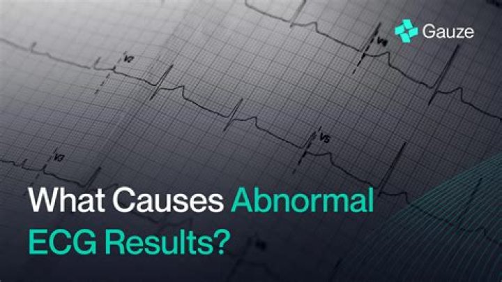
Share on Pinterest An EKG displays P Waves, T Waves, and the QRS Complex. A “normal” EKG is one that shows what is known as sinus rhythm. Sinus rhythm may look like a lot of little bumps, but each relays an important action in the heart.How do you calculate an irregular rhythm?
The second method can be used with an irregular rhythm to estimate the rate. Count the number of R waves in a 6 second strip and multiply by 10. For example, if there are 7 R waves in a 6 second strip, the heart rate is 70 (7x10=70).How do I know if my heart is OK?
Place your index and middle finger of your hand on the inner wrist of the other arm, just below the base of the thumb. You should feel a tapping or pulsing against your fingers. Count the number of taps you feel in 10 seconds. Multiply that number by 6 to find out your heart rate for 1 minute.Can ECG detect heart blockage?
An ECG (electrocardiogram) records the electrical activity of your heart at rest. However, it does not show whether you have asymptomatic blockages in your heart arteries or predict your risk of a future heart attack. The resting ECG is different from a stress or exercise ECG or cardiac imaging test.Can ECG detect stroke?
The following brain and heart tests may be used to help diagnose stroke: Carotid ultrasound, or carotid angiography, shows the insides of the arteries that supply blood to the brain. Electrocardiogram (EKG) is a heart test to help detect heart problems that may have led to a stroke.How accurate is ECG?
The ECG is by far not as accurate as many patients and doctors would like to believe. Often, the findings of a measurement are completely normal even though a heart attack has taken place. As a result, ECG does not detect two out of every three heart attacks at all or not until it is almost too late.Is ECG enough to detect heart problems?
Electrocardiogram (ECG or EKG) to assess the heart rate and rhythm. This test can often detect heart disease, heart attack, an enlarged heart, or abnormal heart rhythms that may cause heart failure. Chest X-ray to see if the heart is enlarged and if the lungs are congested with fluid.Can stress cause heart pain?
If you're often stressed, and you don't have good ways to manage it, you are more likely to have heart disease, high blood pressure, chest pain, or irregular heartbeats. The stress itself can be a problem. Studies also link stress to changes in the way blood clots, which makes a heart attack more likely.What is the best test to check for heart problems?
When heart tests suggest heart disease A test is then needed to pinpoint their location, The gold standard for this purpose is an angiogram taken during cardiac catheterization, an invasive test. A noninvasive alternative is an angiogram taken with an enhanced form of x-ray called computed tomography (CT).What does a ECG show?
Electrocardiogram (ECG) and high blood pressure. An electrocardiogram (ECG) is a test which measures the electrical activity of your heart to show whether or not it is working normally. An ECG records the heart's rhythm and activity on a moving strip of paper or a line on a screen.Can an ECG detect angina?
Angina pectoris or angina is temporary chest pain or discomfort as a result of decreased blood flow to the heart muscle. Your doctor may perform an electrocardiogram (ECG), a stress test without imaging or blood tests to help diagnose your condition.